










Transsylvian Resection of an Insular Low-Grade Glioma Medial to the Porto Alegre Line: Surgical Strategy for Preservation of the Lenticulostriate Arteries. Surgical Video.
Article Sidebar
Main Article Content
Abstract
Introduction: Maximal safe resection remains the primary goal in the surgical management of insular gliomas. However, tumors extending medial to the Porto Alegre Line (PAL) are at increased risk of involving the lenticulostriate arteries (LSAs), making aggressive medial resection potentially devastating. In these cases, preservation of the lenticulostriate arteries rather than subcortical stimulation of the internal capsule represents the critical determinant of the medial limit of resection. We present the surgical strategy for transsylvian resection of an insular low-grade glioma extending beyond the Porto Alegre Line.
Case Report: A 42-year-old woman presented with medically refractory focal epilepsy. Magnetic resonance imaging demonstrated a right insular lesion that was hypointense on T1-weighted imaging, hyperintense on T2-weighted and FLAIR sequences, without gadolinium enhancement. Perfusion MRI demonstrated low relative cerebral blood volume, while MR spectroscopy revealed an elevated choline peak, findings consistent with a diffuse low-grade glioma.Preoperative imaging demonstrated that the medial portion of the tumor extended beyond the Porto Alegre Line, indicating probable intimate relationship with the lenticulostriate arteries. Based on this anatomical finding, a planned subtotal resection was adopted to minimize the risk of permanent motor deficits.
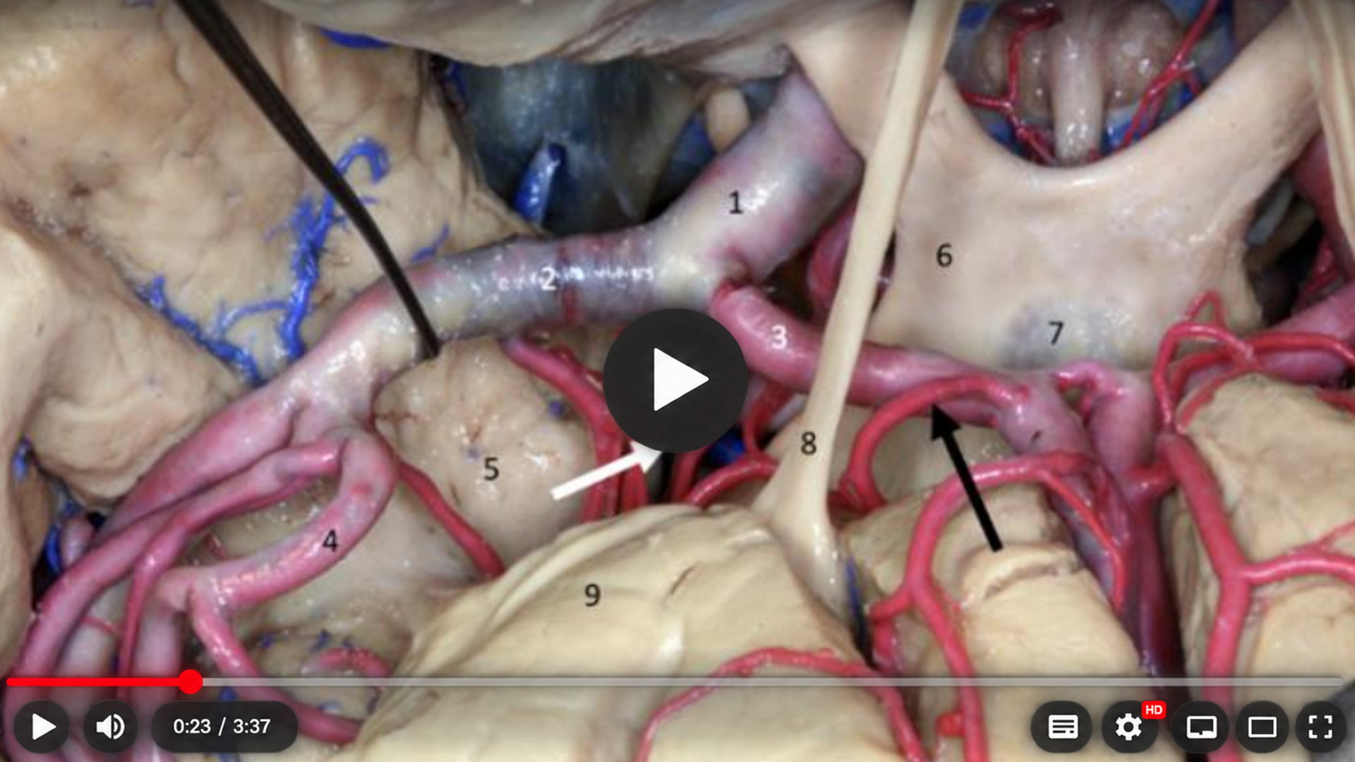
A microsurgical transsylvian approach was performed with wide splitting of the sylvian fissure, allowing exposure of the insular cortex and M2 branches of the middle cerebral artery. Progressive subpial tumor resection was carried out from lateral to medial. Following intraoperative identification of the first lateral lenticulostriate artery, this vessel was deliberately defined as the medial limit of resection, and the tumor surrounding the lenticulostriate complex was intentionally preserved. In this surgical strategy, the anatomical location of the lenticulostriate arteries, rather than subcortical stimulation of the internal capsule, was considered the principal determinant of the safe medial resection boundary.
Histopathological examination confirmed a diffuse low-grade glioma.
Results: The patient experienced no new postoperative neurological deficits. At follow-up, she remained seizure-free, with complete resolution of her medically refractory epilepsy.
Conclusion: The Porto Alegre Line is a practical and reliable preoperative anatomical landmark for predicting medial extension of insular gliomas and their relationship with the lenticulostriate arteries. In tumors extending medial to this line, planned preservation of the lenticulostriate artery complex should take precedence over pursuit of gross-total resection, as these perforating vessels - not the internal capsule identified by subcortical stimulation - constitute the true functional limit of safe medial resection. This strategy maximizes neurological preservation while maintaining the oncological principles of maximal safe resection.
Article Details
Section

This work is licensed under a Creative Commons Attribution 4.0 International License.
References
1. Isolan GR, Bianchin MM, Bragatti JA, Torres C, Schwartsmann G. Musical hallucinations following insular glioma resection. Neurosurg Focus. 2010;28(2):E9. https://doi.org/10.3171/2009.12.FOCUSFOCUS09243
2. Isolan GR, Buffon V, Maldonado I, Monteiro JM, Yağmurlu K, Ribas CAPM, et al. Avoiding vascular complications in insular glioma surgery: a microsurgical anatomy study and critical reflections regarding intraoperative findings. Front Surg. 2022;9:906466. https://doi.org/10.3389/fsurg.2022.906466
3. Isolan GR, Marth V, Frizon L, Dini L, Dini S, Yamaki VN, et al. Surgical treatment of drug-resistant epilepsy caused by gliomas in eloquent areas: experience report. Arq Neuropsiquiatr. 2019;77(11):797-805. https://doi.org/10.1590/0004-282X20190160
4. Isolan GR, Bark SA, Monteiro JM, Mattei TA, Yağmurlu K, Gonçalves RF, et al. Porto Alegre Line predicts lenticulostriate arteries encasement and extent of resection in insular gliomas: a preliminary study. Front Surg. 2025;12:1414302. https://doi.org/10.3389/fsurg.2025.1414302
5. Isolan GR, Campero A, Ajler P, Farina EMG, Frigeri TM, Dini LI. Parte I: anatomía microquirúrgica tridimensional de la ínsula. Rev Argent Neuroc. 2020;34(2):100-115.
6. Buffon VA, Isolan GR, Barbosa B, Brito JNPO, de Araújo RL, Almeida TAL. Gliomas da ínsula: revisão anatômica e sua correlação com a extensão da ressecção, morbidade e sobrevida. BioSCIENCE. 2024;82:e00007. https://doi.org/10.55684/2024.82.e00007
7. Isolan GR. Tumores do cérebro e da base do crânio: do laboratório de microanatomia neurocirúrgica à sala de cirurgia. 1st ed. Rio de Janeiro: Thieme Revinter; 2026.
8. Isolan GR, Bark SA, Buffon V, Dini LI, Becker LG, Malafaia O, et al. As sete regras para obter sucesso na cirurgia dos gliomas da ínsula. BioSCIENCE. 2024;82:e00066. https://doi.org/10.55684/2024.82.e00066
9. Isolan GR, Antunes ACM, Dini LI, Stefani MA, da Costa CRF, Falcetta F, et al. Insular gliomas: general aspects and surgical technique. J Bras Neurocirurg. 2008;19(4):7-13. https://doi.org/10.22290/jbnc.v19i4.689
10. Vanacôr CN, Isolan GR, Yu YH, Telles JPM, Oberman DZ, Rabelo NN, et al. Microsurgical anatomy of language. Clin Anat. 2021;34(1):154-168. https://doi.org/10.1002/ca.23681
11. Isolan GR, Rodrigues EM, Marques VMO, Becker LG, Dini LI. Conceitos técnicos para evitar a lesão das radiações ópticas na abordagem para a região mesial do lobo temporal. J Bras Neurocirurg. 2021;32(1):29-49. https://doi.org/10.22290/jbnc.v32i1.1940
12. Isolan GR, Marrone ACH, Marrone LCP, Stefani MA, da Costa JC, Telles JPM, et al. Vascularization of the uncus: anatomical study and clinical implications. Surg Neurol Int. 2021;12:393. https://doi.org/10.25259/SNI_616_2021
13. Isolan GR, Campero A, Ajler PM, Farina EM, Frigeri TM, Dini LI. El lóbulo de la ínsula: Parte 2 - anatomía microquirúrgica y correlación clínico-quirúrgica. Surg Neurol Int. 2020;11(Suppl 1):S37-S52. https://doi.org/10.25259/SNI_679_2020
14. Isolan GR, Stefani MA, Schneider FL, Claudino HA, Yu YH, Choi GG, et al. Hippocampal vascularization: proposal for a new classification. Surg Neurol Int. 2020;11:378. https://doi.org/10.25259/SNI_708_2020
15. Bark SA, Isolan GR, Barbosa B, Brito JNPO, de Araújo RL, Almeida T. As artérias lenticuloestriadas podem interferir na extensão da ressecção tumoral e sobrevida nos gliomas da ínsula? BioSCIENCE. 2023;81(2):108-119. https://doi.org/10.55684/81.2.20
16. Isolan GR. CEANNE - Igualando a qualidade de atendimento neurocirúrgico oncológico entre pacientes atendidos nos sistemas de saúde público e privado em hospitais brasileiros. BioSCIENCE. 2024;82(Suppl 1):e002. https://doi.org/10.55684/2024.82.S1.e002
17. Isolan GR, Santos RS, Badalotti R, Rutz LEAC, Caron VF, Gonçalves RF, et al. O conceito de área de Broca não existe em gliomas de baixo grau – uma série de casos. BioSCIENCE. 2024;82(Suppl 1):e003. https://doi.org/10.55684/2024.82.S1.e003