










En Plaque Sphenoid Wing Meningioma: How I Do It? Surgical Video.
Article Sidebar
Main Article Content
Abstract
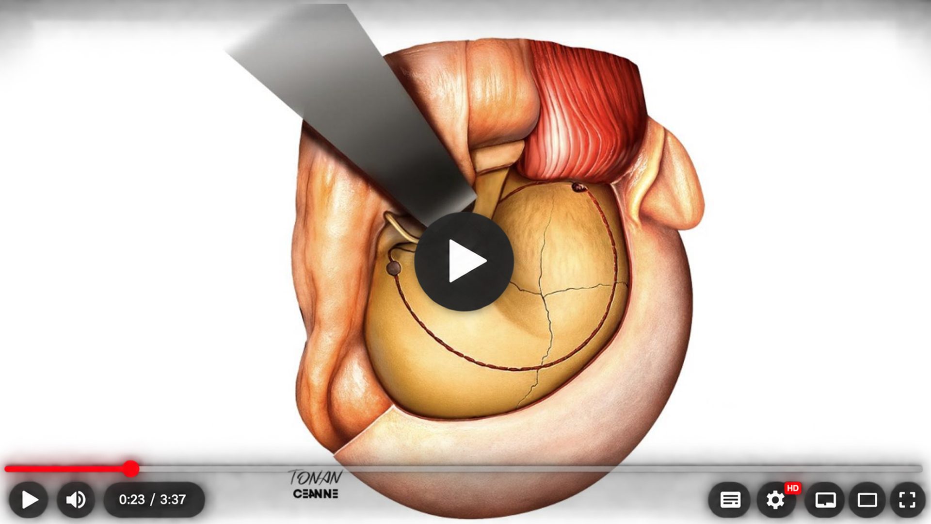
Introduction: En plaque sphenoid wing meningiomas are uncommon skull base tumors characterized by diffuse dural infiltration associated with extensive hyperostosis of the sphenoid wing and adjacent cranio-orbital bones. Progressive proptosis is the most frequent presenting symptom and is primarily caused by tumoral bone invasion rather than soft tissue compression. Radical treatment requires complete removal of both the dural lesion and the infiltrated bone to minimize recurrence. This surgical video demonstrates our microsurgical technique for the management of these challenging cranio-orbital tumors.
Case Report: A 37-year-old woman presented with progressive right-sided proptosis without visual impairment. Neurological and ophthalmological examinations demonstrated preserved visual acuity, intact visual fields, and normal extraocular movements. Computed tomography revealed marked hyperostosis involving the right sphenoid wing, anterior clinoid process, lateral orbital wall, orbital roof, and pterygoid plate. Contrast-enhanced magnetic resonance imaging demonstrated an en plaque sphenoid wing meningioma with diffuse dural thickening and extension into the lateral orbit, producing significant orbital compression. A right pterional craniotomy was performed with removal of the superior and lateral orbital walls to achieve a wide cranio-orbital exposure. After microsurgical devascularization of the lesion, complete resection of the en plaque meningioma and its dural attachment was achieved. Extensive drilling of all hyperostotic bone, including the anterior clinoid process, sphenoid wing, lateral orbital wall, orbital roof, and pterygoid plate, was performed until normal cancellous bone was encountered. The orbital walls were reconstructed at the conclusion of the procedure.
Results: The postoperative course was uneventful, with preservation of visual function and no new neurological deficits. The patient's proptosis progressively resolved, resulting in excellent cosmetic and functional outcomes. At 10 years of clinical and radiological follow-up, there was no evidence of tumor recurrence.
Conclusion: Complete microsurgical resection of en plaque sphenoid wing meningiomas requires meticulous removal of both the dural tumor and the involved cranio-orbital bone. A pterional cranio-orbital approach provides excellent exposure for radical resection while preserving visual function. This case demonstrates that aggressive removal of hyperostotic bone can achieve durable long-term tumor control with excellent functional and cosmetic results.
Article Details
Section

This work is licensed under a Creative Commons Attribution 4.0 International License.
References
1. Al-Mefty O. Clinoidal meningiomas. J Neurosurg. 1990;73(6):840-849. https://doi.org/10.3171/jns.1990.73.6.0840
2. Simon M, Schramm J. Sphenoid wing meningiomas. In: Pamir MN, Black PM, Fahlbusch R, editors. Meningiomas: a comprehensive text. Philadelphia: Saunders Elsevier; 2010. p. 427-443. https://doi.org/10.1016/B978-1-4160-5654-6.00032-5
3. Nakasu S, Fukami T, Jito J, Nozaki K. Recurrence and regrowth of benign meningiomas. Brain Tumor Pathol. 2009;26(2):69-72. https://doi.org/10.1007/s10014-009-0251-2
4. De Jesús O, Sekhar LN, Parikh HK, Wright DC, Wagner DP. Long-term follow-up of patients with meningiomas involving the cavernous sinus: recurrence, progression, and quality of life. Neurosurgery. 1996;39(5):915-920. https://doi.org/10.1097/00006123-199611000-00005
5. Rhoton AL Jr. The orbit. Neurosurgery. 2002;51(4 Suppl):S303-S334. https://doi.org/10.1097/00006123-200210001-00008
6. Dolenc VV. Anatomy and surgery of the cavernous sinus. Vienna: Springer-Verlag; 1989. https://doi.org/10.1007/978-3-7091-6942-1
7. Al-Mefty O. Operative atlas of meningiomas. Philadelphia: Lippincott-Raven; 1998.
8. Ouyang T, Zhang N, Wang L, Li Z, Chen J. Sphenoid wing meningiomas: surgical strategies and evaluation of prognostic factors influencing clinical outcomes. Clin Neurol Neurosurg. 2015;134:85-90. https://doi.org/10.1016/j.clineuro.2015.04.016
9. Yaşargil MG. Microneurosurgery. Volume IVB: microneurosurgery of CNS tumors. Stuttgart: Thieme; 1996.
10. Isolan GR. Tumores do cérebro e da base do crânio: do laboratório de microanatomia neurocirúrgica à sala de cirurgia. 1st ed. Rio de Janeiro: Thieme Revinter; 2026.
11. Cândido DNC, Monteiro JM, Barbosa B, Brito JNPO, de Araújo RL, Almeida TAL. O papel do forame clinocarotídeo na cirurgia de aneurismas da artéria oftálmica. BioSCIENCE. 2024;82(1):e005. https://doi.org/10.55684/2024.82.e005
12. Silveira GCP, Gonçalves RF, Tomasi G, Saciloto B, Martins LML, Nogueira GN, et al. Alterações neuropsiquiátricas em meningeoma petroclival. BioSCIENCE. 2024;82(S1):e029. https://doi.org/10.55684/2024.82.S1.e029