










Pretemporal Pterional Approach for Sphenopetroclivial Meningioma Causing Trigeminal Neuropathy: Surgical Video
Article Sidebar
Main Article Content
Abstract
Introduction: Sphenopetroclival meningiomas represent a major surgical challenge because of their intimate relationship with critical neurovascular structures, particularly the cavernous sinus and cranial nerves. Complete resection may be limited by the risk of permanent cranial nerve injury; therefore, selective decompression of symptomatic neural structures followed by radiosurgery for residual tumor has become an effective treatment strategy for cavernous sinus meningioma causing trigeminal neuropathy. The pretemporal pterional approach provides wide exposure of cisternal and cavernous sinus components while minimizing brain retraction.
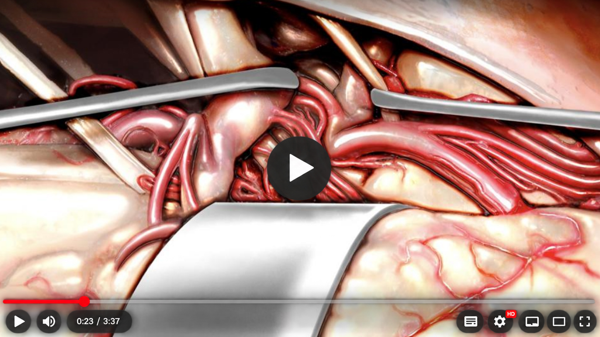
Case Report: A 33-year-old woman presented with refractory neuropathic pain in the right trigeminal nerve distribution. Contrast-enhanced MRI demonstrated a small sphenopetroclival meningioma with cisternal and intracavernous components. Neurological examination revealed no cranial nerve palsies or extraocular muscle deficits other than trigeminal neuropathy. Because of debilitating pain refractory to conservative management, surgery was indicated to decompress the trigeminal nerve. A pretemporal pterional approach was performed with wide dissection of the Sylvian fissure. Bridging veins from the temporal pole to the sphenoparietal sinus were coagulated and sacrificed to allow gentle temporal lobe retraction. This provided visualization of the right internal carotid artery and the oculomotor and trochlear nerves at the tentorial incisura. After coagulation and sectioning of the tentorial incisura, the cisternal tumor component was removed, achieving trigeminal nerve decompression. The intracavernous component was intentionally left in situ to avoid cranial nerve injury.
Results: Postoperative imaging confirmed successful trigeminal nerve decompression with residual intracavernous tumor. Two years after surgery, growth of the residual component was documented, and the patient was referred for stereotactic radiosurgery. After 14 years of follow-up, no further tumor growth was observed, and the patient remained free of trigeminal symptoms.
Conclusion: This case supports a staged strategy combining pretemporal pterional microsurgical decompression of symptomatic neural structures with radiosurgery for residual cavernous sinus meningioma. The approach provides wide exposure of the suprasellar, interpeduncular, and petroclival regions and enables selective resection of accessible tumor while preserving critical neurovascular structures. Radiosurgery can provide durable control of intentionally retained intracavernous disease, reducing the risk associated with aggressive microsurgical resection.
Article Details
Section

This work is licensed under a Creative Commons Attribution 4.0 International License.
References
1. Al-Mefty R, Dunn IF, Pravdenkova S, Abolfotoh M, Al-Mefty O. True petroclival meningiomas: results of surgical management. J Neurosurg. 2014;120(1):40-51. https://doi.org/10.3171/2013.8.JNS13535
2. Al-Mefty O. Overview of petroclival meningiomas. In: Al-Mefty O, editor. Meningiomas. Philadelphia: Elsevier; 2010. p. 477-486.
3. Al-Mefty O, Smith RR, Fox JL. Petrosal approach for petroclival meningiomas. Neurosurgery. 1988;22(3):510-517. https://doi.org/10.1227/00006123-198803000-00010
4. Aum D, Rassi MS, Al-Mefty O. Petroclival meningiomas and the petrosal approach. In: Badie B, editor. Neurosurgical Operative Atlas: Neuro-Oncology. Amsterdam: Elsevier; 2020. p. 133-141.
5. Isolan GR, Wayhs SY, Lepski GA, Dini LI, Lavinsky J. Petroclival meningiomas: factors determining the choice of approach. J Neurol Surg B Skull Base. 2018;79(4):367-378. https://doi.org/10.1055/s-0037-1608654.
6. Isolan GR, Wayhs SY, Araújo RL, Aguiar PHP, Reyes Pinto JA, Marques VMO. Surgical approaches to petroclival meningiomas. Part 1: microsurgical anatomy. Surg Neurol Int. 2022;13:277. https://doi.org/10.25259/SNI_413_2022.
7. Isolan GR, Lavinsky J, Marques VMO, Monteiro JM, Santos RS, Aguiar PHP. Surgical approaches to petroclival meningiomas. Part 2: narrative review of what we learned with 30 cases. Surg Neurol Int. 2022;13:515. https://doi.org/10.25259/SNI_696_2022.
8. Isolan GR, Krayenbühl N, de Oliveira E, Al-Mefty O. Microsurgical anatomy of the cavernous sinus: measurements of the triangles in and around it. Skull Base. 2007;17(6):357-367. https://doi.org/10.1055/s-2007-985194.
9. Isolan GR, Braga F, Araújo RL. Cavernous sinus triangles: correlation between cranial and endonasal visualization. Arq Bras Neurocir. 2021;40(3):e295-e296. https://doi.org/10.1055/s-0041-1733996.
10. Isolan GR. Tumores do cérebro e da base do crânio: do laboratório de microanatomia neurocirúrgica à sala de cirurgia. 1ª ed. Rio de Janeiro: Thieme Revinter; 2026. 812 p.
11. Balestri HP, Batista FS, Cardoso CEP, Silva IS, Rutz LEAC, Campos ECR, et al. Acesso pré-temporal para meningeoma esfenopetroclival. BioSCIENCE. 2024;82(S1):e004. https://doi.org/10.55684/2024.82.S1.e004.
12. Lavinsky J, Nogueira GN, Gonçalves RF, Schuster AK, Roesler R, de Andrade JMC, et al. O risco de radiocirurgia como tratamento primário para tumores supostamente benignos da base do crânio. BioSCIENCE. 2024;82(S1):e016. https://doi.org/10.55684/2024.82.S1.e016.